In vitro Wound Healing Potential and Identification of Bioactive Compounds from Aegle marmelos and Mucuna pruriens Methanolic Extract
Volume 1, Issue 1, Pages 17-28
History
Received 06 November 2025
Revised 22 November 2025
Accepted 04 December 2025
Keywords
Aegle marmelos, Mucuna pruriens, Wound healing, CAM assay, Bioguided fractionation.
Open Access
This is an open access article under the CC BY license https://creativecommons.org/licenses/by/4.0/.
RESULTS AND DISCUSSION
The method developed for HPLC fingerprinting provided a quick analysis of the isolated fraction. The components gallic acid, rutin and quercitin were identified by comparing retention times, with the retention times of gallic acid, rutin and quercitin standards. The conditions used led to a good separation of the peaks which could be identified in the chromatogram as gallic acid (Rt = 9.9), rutin (Rt = 15.9) and quercitin (Rt = 20.6). The chromatograms are shown in Figures 2, 4 and 6. They were identified by comparison with the chromatogram of the reference compounds obtained under the same conditions and the respective UV spectra, obtained on line. The chromatograms are shown in Figures 1, 3 and 5.
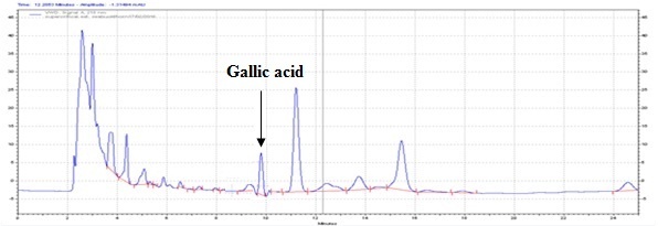
Figure 2 - HPLC chromatogram of isolated fraction AM-3-03.
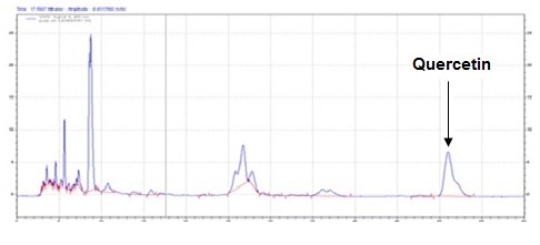
Figure 4 - HPLC chromatogram of isolated fraction AM-4-01-01.
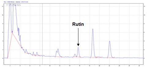
Figure 6 - HPLC chromatogram of isolated fraction MP-4-03-01-01-01.
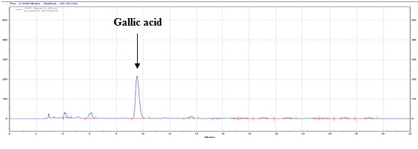
Figure 1 - HPLC chromatogram of standard gallic acid.
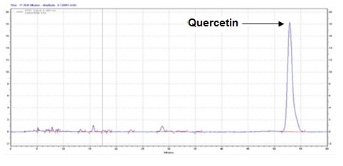
Figure 3 - HPLC chromatogram of standard quercitin.
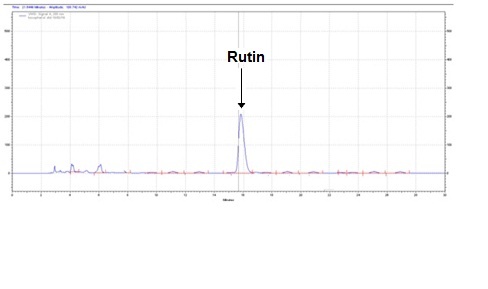
Figure 5 - HPLC chromatogram of standard rutin.
The identified peaks were considered to represent only one compound each because the UV spectra at the upslope and down slope inflection points, in each peak, were indistinguishable. The analysis time is an important factor in analytical work and the run time should be reduced to a minimum in order to optimize equipment use and reduce solvent consumption. Although the peak of gallic acid, rutin and quercitin were well resolved under the conditions used for the fingerprinting chromatogram, when we increased the organic modifier content of the mobile phase the peaks of interest were still well resolved but with reduced retention time. The described HPLC procedure could be useful for the qualitative and quantitative analysis of flavonoids in plant materials.
Isolated fraction AM-3-03 was found to be light yellowish in color, melting point 260–262°C and observed yellowish in colour under UV light. Soluble in methanol, ethanol and water, identified as that the compound may be derivative of gallic acid (Figures 7–10) as interpreted from the different spectral data (Table 2).
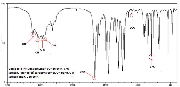
Figure 7 - Showing Infra red spectrum (IR) of isolated fraction (AM-3-03).
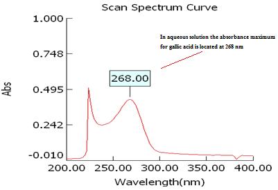
Figure 8 - Showing UV-Vis spectra of isolated fraction (AM-3-03).
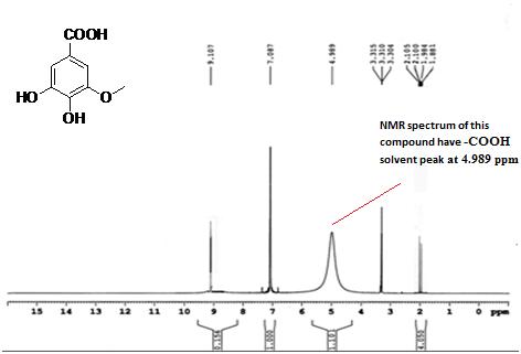
Figure 9 - Showing H NMR of isolated fraction (AM-3-03).
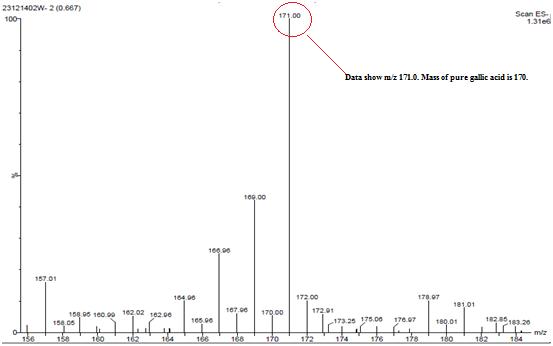
Figure 10 - Showing MASS spectra of isolated fraction (AM-3-03).
| Method | Spectral interpretation |
|---|---|
| UVmax | 268 nm |
| IR | Stretching band at 3259.95 cm-1 (s) due to OH stretching; at 3015.23 (s) due to Aromatic CH str.; 2932.28, 2851.91 due to Aliphatic C-H str.; 1704.91due to C=O str.; 71.34, 1072.6 due to C-O str., 1651.08, 1503.42, 1417.91 due to Aromatic C=C; 775.83 due to Aromatic C-H out of plane |
| 1HNMR (ppm) | 7.087 ppm due to s, 2H, ArH; at 9.107 ppm due to H s, 1H, -COOH; at 4.989 ppm due to s, 3H, 3 OH |
| MASS (m/z %) | 171.0 (due to molecular ion) |
| Structure | 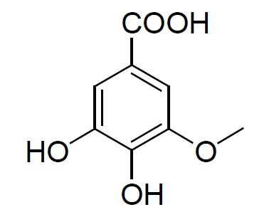 3,4-dihydroxy-5-methoxybenzoic acid |
Isolated fraction AM-4-01-01 was found to be pale yellow powder, melting point 311–315°C and observed yellowish in colour under UV light. Soluble in ethanol, methanol, chloroform, DMSO and partially insoluble in water, identified as that the compound maybe derivative of quercetin (Figures 11–14) as interpreted from the different spectral data (Table 3).
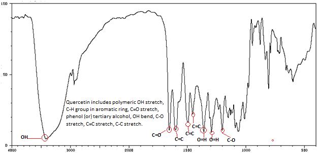
Figure 11 - Showing IR spectrum of isolated fraction (AM-4-01-01).
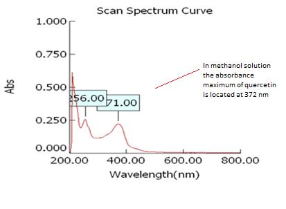
Figure 12 - Showing UV-Vis spectra of isolated fraction (AM-4-01-01).
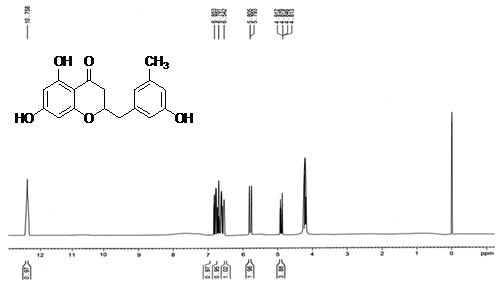
Figure 13 - Showing H NMR of isolated fraction (AM-4-01-01).
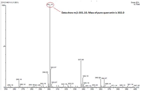
Figure 14 - Showing MASS spectra of isolated fraction (AM-4-01-01).
| Method | Spectral interpretation |
|---|---|
| UVmax | 256 nm and 271 nm |
| IR | 3495.12 (OH, stretch), 1696.43 (C=O, stretch), 1610.55, 1537.28, 1427.20 (C=C stretch), 1309.07 (OH, In-plane bend), 1246-1199 (C-O stretch), 1023.81 (OH, Out-plane bend), 864.49 (Ar-H, Out of plane bend). |
| 1HNMR (ppm) | 8.04 (m, 2H, Ar-H), 7.95–7.96 (multiplat, Ar-H), 6.37-6.38 (m, 1H Al-OH), 6.17-6.20 (m, 1H, Ar-OH), 5.22-5.24 (m, 2H, Ar-OH), 4.47 (m, 1H, Ar-OH). |
| MASS (m/z %) | 301.2 (Base peak) |
| Structure | 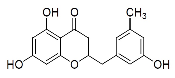 2-(3-hydroxy-5-methylbenzyl)-2,3-dihydro-5,7-dihydroxychromen-4-one |
Isolated fraction MP-4-03-01-01-01 was found to be light yellow powder, melting point 311–242–244°C and observed yellowish in colour under UV light. Soluble in ethanol, methanol, chloroform, DMSO and partially insoluble in water, identified as that the compound maybe derivative of rutin (Figures 15–18) as interpreted from the different spectral data (Table 4).
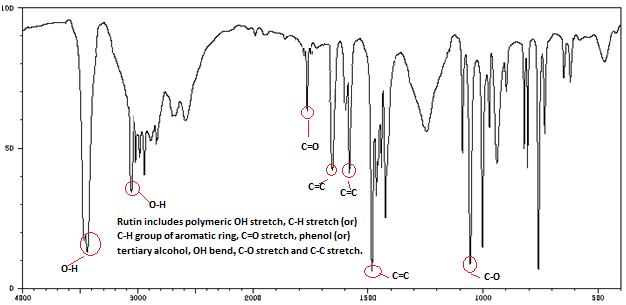
Figure 15 - Showing IR spectrum of isolated fraction (MP-4-03-01-01-01).
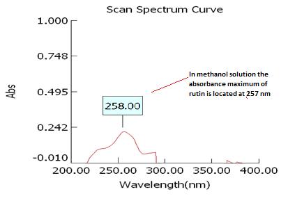
Figure 16 - Showing UV-Vis spectra of isolated fraction (MP-4-03-01-01-01).
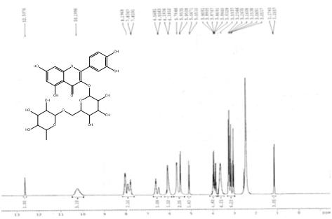
Figure 17 - Showing H NMR of isolated fraction (MP-4-03-01-01-01).
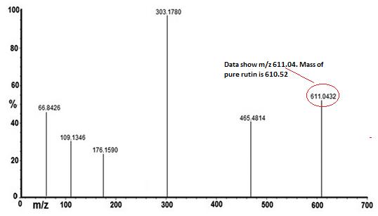
Figure 18 - Showing MASS spectra of isolated fraction (MP-4-03-01-01-01).
| Method | Spectral interpretation |
|---|---|
| UVmax | 258 |
| IR | C-O-1076.58, C=C-1667.18 & 1473.29, C=O-1729.29, O-H-3423.06 & 3140.24, CH2-OH-1573.25 |
| 1HNMR (ppm) | OH-12.5976 (s, 1H), OH-10.1996 (s, 4H), CH-8.1968 (s, 1H), CH-7.8367 (d, 1H), CH-6.568 (d, 1H), CH-6.1936 (d, 1H), CH-5.9666 (s, 1H), CH2-OH-5.9535 (d, 2H), CH-5.0671 (d, 1H), CH-3.9081-3.8329 (p, 4H), OH-3.5937 (s, 6H), CH-3.2566-3.0517 (triple triplet, 6H), CH3-1.1740 (d, 3H) |
| MASS (m/z %) | 611.04-M scan |
| Structure |  C27H30O16 |
Isolated fractions at different concentrations, along with VEGF as a positive control and DMSO as a negative control, was tested in the CAM assay for its angiogenic activity and it was demonstrated that the CAM assay are versatile tools for the in vitro evaluation of small quantities of natural compounds or isolated compounds, enabling the detection of possible angiogenic or antiangiogenic effects of the test compound to evaluate its property as an angiogenic drug candidature.
After incubation period (Figure 19), VEGF treatment led to a simulation with a strong network of sprouting capillaries and increased vessel density within the ring and treatment with isolated fractions at different concentrations showed the formation of capillaries similar to the VEGF. No changes in vascular structure or density were observed with DMSO. CAM implanted with VEGF showed a significant increase of new blood vessels in the VEGF control group suggests that the added VEGF was the only factor which could have stimulated the ectopic angiogenesis in CAM.
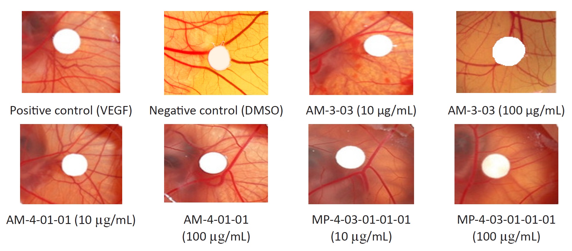
Figure 19 - Images of CAMs implanted with VEGF and different concentrations of isolated fractions.
CAM implanted with isolated fractions AM-3-03, AM-4-01-01 and MP-4-03-01-01-01 at 10 µg/mL concentration inhibited angiogenesis by 62.40 ± 8.89%, 46.68 ± 4.51% and 68.35 ± 4.07% respectively, while the higher concentrations of 10 µg/mL showed a moderate degree of angiogenesis inhibition of, by 60.00 ± 5.74%, 28.26 ± 1.58% and 61.14 ± 7.86%, respectively (Table 5). Negative control (DMSO) treatment revealed a smaller number of newly formed blood vessels (8.75 ± 2.28) compared to new blood vessels (36.00 ± 5.58) in positive con-trol (VEGF).
| Treatment | Concentration of extract (µg/mL) | Number of new blood vessels | Angiogenic inhibition (%) |
|---|---|---|---|
| Positive control (VEGF) | 36.00 ± 5.58 | 0 | |
| Negative control (DMSO) | 8.75 ± 2.28 | 0 | |
| AM-3-03 | 10 | 14.25 ± 3.81 | 62.40 ± 8.89 |
| 100 | 17.50 ± 2.79 | 60.00 ± 5.74 | |
| AM-4-01-01 | 10 | 19.25 ± 3.25 | 46.68 ± 4.51 |
| 100 | 26.50 ± 1.02 | 28.26 ± 1.58 | |
| MP-4-03-01-01-01 | 10 | 10.75 ± 1.75 | 68.35 ± 4.07 |
| 100 | 15.50 ± 4.96 | 61.14 ± 7.86 |
Angiogenesis is important in normal process such as development of embryo formation of corpus luteum and wound healing [22]. Angiogenesis during wound repair serves the dual function of providing the nutients demanded by the healing tissues and contributing to structural repair through the formation of granulation tissue [23]. The chicken CAM assay revealed a reduction in the antiangiogenic effect. VEGF has endothelial cell-specific mitogenic activity and stimulates angiogenesis in vivo and in vitro [24]. VEGF is an important pro-angiogenic cytokine and improves angiogenesis during wound healing by stimulating the migration and proliferation of endothelial cells through the extra cellular matrix [25]. The methanolic extract being the most bioactive crude extract suggests fully that the compound(s) responsible for the proliferative action may be chemically polar and inorganic because methanol is an inorganic solvent in addition to its polar property. These active compounds belong to the group of flavonoid compounds, this group of compounds is known to be beneficial in wound healing treatment [26]. This potential could be due to the ability to regulate any of the proteins or chemotactic factors involved in the healing process at the molecular level. The evaluation of the molecular mechanism behind this potency requires further studies in the established model which can be extended to wounds of different types and severity. The identified bioactive compounds in the methanolic crude extract of Aegle marmelos and Mucuna pruriens may serve as a lead in the drug discovery and development of a wound healing agent that may enhance wound healing.
Conflict of Interest
The authors declare that they have no conflicts of interest to disclose.
Author’s Contribution
Fedelic Ashish Toppo performed the research work, and wrote the manuscript with support from Mahendra C Gunde. Mahendra C Gunde verified the assisted with data interpretation. Both authors discussed the results and approved the final manuscript.